- More Than Teeth
- Posts
- Part 2 of 3: From Sleep Study to Treatment Plan
Part 2 of 3: From Sleep Study to Treatment Plan
Where Dentistry Meets Whole-Body Health Michael Bennett, DDS, PhD & Cathy Bennett, MS, NBCHWC
This is More Than Teeth. The newsletter that helps dental sleep professionals get 1% better every week.
Good Morning.
Last week we mapped the territory. This week, we hand you the compass.
Part 1 was the why. Part 2 is the how — read the sleep study, identify the phenotype, build the augmentation plan, and know which therapies are in dental scope versus which require a phone call to your sleep medicine partner.
Key Takeaways
If you've only got 30 seconds, here's the whole issue:
Four PSG metrics tell you the phenotype: positional AHI breakdown, REM vs. NREM AHI, arousal index relative to AHI, and pattern of desaturation.
Each PALM phenotype has a primary therapy. Anatomical → OAT. Arousal threshold → pharmacologic (referral). Loop gain → oxygen or acetazolamide (referral). Muscle responsiveness → myofunctional therapy ± hypoglossal nerve stimulation.
Combination is the norm. Most patients are a dominant phenotype plus one or two contributors. Plan the augmentation, don't just prescribe a device.
Audit the nose first. Tier 1 (saline + xylitol + dilator strips) is within your scope and should be performed on every patient before you blame the appliance.
Mouth taping requires a patent nose. No exceptions.
7-minute read 👇 (deep dive below)
Reading the PSG for Phenotype
A note before the worksheet: most of your OAT candidates will walk in with a home sleep test (HST), often a ring-based cardiopulmonary coupling (CPC) study like SleepImage, rather than a full in-lab PSG. The framework below is built on PSG data because it provides the most complete picture and the clearest teaching reference. If you're reading HST output instead, the four questions still work — the data fields are just different. See the HST/CPC companion worksheet linked at the end of this issue for the adapted version.
Most sleep study reports provide more than enough information to phenotype a patient. The problem isn't data, it's knowing which four numbers actually matter for an OAT case selection decision. Here they are.
1. Positional AHI breakdown (supine vs. lateral). Supine AHI more than two times lateral AHI is the strongest single signature for a predominantly anatomical phenotype. These are your highest-percentage OAT candidates. If the report doesn't break it out, the technician's narrative usually mentions it.
2. REM vs. NREM AHI. REM-predominant disease (REM AHI substantially higher than NREM AHI, often with deeper desaturations during REM) points away from anatomical factors and toward muscle responsiveness and loop-gain phenotypes. REM is when the pharyngeal dilator tone drops out, and respiratory control becomes twitchy. A patient whose AHI triples in REM is telling you the appliance alone may not be enough.
3. Arousal index relative to AHI. A high arousal index (>25/hr) paired with a relatively modest AHI and preserved oxygen nadir (>85%) is a low-arousal-threshold signature. These patients wake up at the first whiff of resistance, which fragments sleep and perpetuates the cycle. Mandibular advancement won't change how their brain responds to airway loading.
4. Pattern of desaturation. Cyclical, periodic breathing — a metronomic up-and-down of SpO2 across the night — flags high loop gain. Deep, isolated REM desaturations flag muscle responsiveness. Position-locked desaturations that resolve when the patient turns onto their side flag anatomy. Look at the hypnogram, not just the summary stats.
Map those four metrics onto the PALM model, and the dominant phenotype usually declares itself.
Most patients aren't pure phenotypes — they're a dominant trait plus one or two contributing traits. That's a feature, not a bug. The phenotyping tells you what to combine, not just what to prescribe.

The Augmentation Playbook
For each phenotype, here's what the evidence supports — split by what stays on your side of the wall and what needs your sleep medicine partner.
Anatomical (P)
Primary therapy: Oral appliance therapy. This is where OAT was designed to shine.
In dental scope:
Properly titrated MAD with documented advancement
Positional therapy device or supine-sleep avoidance training for the supine-dominant subset
Confirm and maintain nasal patency (see protocol below)
Combination MAD + CPAP for the severe-baseline subset who tolerate it
Co-management: Severe baseline AHI (>30) with significant residual disease after titration. Time for a combination conversation.
Arousal Threshold (A)
Primary therapy: Raising the arousal threshold — pharmacologically, behaviorally, or both. The MAD addresses the anatomical contribution; it doesn't change cortical reactivity.
In dental scope:
Sleep hygiene counseling (caffeine cutoff, alcohol within 3 hours of bed, screen exposure)
Optimize OAT to handle the structural component
Document and refer to the arousal-threshold contribution
Co-management (sleep medicine scope):
Trial of an arousal-threshold-raising agent. Eszopiclone has the most direct evidence (Edwards et al., 2016 — eszopiclone raised arousal threshold and reduced AHI in low-threshold patients).
Trazodone is sometimes used off-label for a similar effect.
This is a prescription conversation, not a dental one — but identifying the phenotype is what gets the patient into that conversation.
Loop Gain (L)
Primary therapy: Stabilizing the respiratory control loop. Mechanical advancement doesn't reach the chemoreflex.
In dental scope:
Optimize OAT to reduce respiratory load
Optimize nasal patency to reduce ventilatory variability
Recognize and refer
Co-management (sleep medicine scope):
Supplemental low-flow oxygen (Wellman et al., 2008 — oxygen reduced AHI in high-loop-gain patients).
Acetazolamide (Edwards et al., 2012 — reduced loop gain and lowered AHI in selected patients).
Both require a sleep physician's prescription and monitoring.
Muscle Responsiveness (M)
Primary therapy: Building or replacing pharyngeal dilator tone.
In dental scope:
Myofunctional therapy referral — well-supported as an adjunct (Camacho et al., 2015 meta-analysis: 50% AHI reduction in adults, 62% in children).
A combination of MAD + myofunctional therapy is showing promise in the literature.
Address daytime habits: chronic mouth breathing, tongue posture.
Co-management:
Hypoglossal nerve stimulation (Inspire) for the right candidate — the STAR trial (Strollo et al., 2014, NEJM) remains the foundational evidence; FDA-approved for selected moderate-severe patients who fail CPAP. Sleep medicine and ENT co-manage; you are the referring identifier.
A note on this issue's sponsor: More Than Teeth is sponsored in part by Xlear, a Utah-based company whose xylitol-based saline products we use as our Tier 1 daily-use nasal protocol at Advanced Dental Care. Xylitol's effect on nasal mucosal flora makes it a clinically meaningful upgrade over plain saline for the chronic mouth-breathing patients who otherwise quietly undermine OAT outcomes.
The Tiered Nasal Protocol
Before you escalate anything, audit the nose. Nasal obstruction undermines every phenotype's response to mandibular advancement, and most OAT non-responders have something nasal going on that nobody asked about.
Tier 1 — Conservative (dental scope)
Saline irrigation. High-volume sinus rinse or neti pot, 1–2x daily. Strongest evidence base of any nasal intervention for chronic congestion (Hermelingmeier et al., 2012, meta-analysis).
Xylitol-based nasal spray. A meaningful upgrade over plain saline for chronic mouth-breathers — xylitol disrupts bacterial adhesion to nasal mucosa, which addresses one of the inflammatory drivers of chronic congestion. Xlear is the category-leading product in this space and the one we use at Advanced Dental Care; we recommend it as the daily-use option after the morning rinse. Recommended dosing: Xlear Nasal Spray, 2–3 sprays per nostril 1–2x daily, or Xlear MAX for patients with significant chronic congestion.
Nasal dilator strips at night (Breathe Right and similar). Mechanical, cheap, low-risk, useful trialable adjunct.
Positional and environmental. Bedroom allergen reduction, head-of-bed elevation, side-sleeping.
Tier 2 — Pharmacologic (PCP / sleep med scope; you recommend)
Intranasal corticosteroid (fluticasone, mometasone) — 6-week trial for inflammatory/allergic component.
Second-generation antihistamines if allergic rhinitis is suspected.
Avoid chronic decongestants — rebound congestion makes things worse over weeks.
Tier 3 — ENT referral
Deviated septum, hypertrophied turbinates, collapsed nasal valves, adenoid remnants in adults — all surgical territory.
Refer when conservative and pharmacologic measures have had a fair trial (8–12 weeks) and mouth breathing persists.
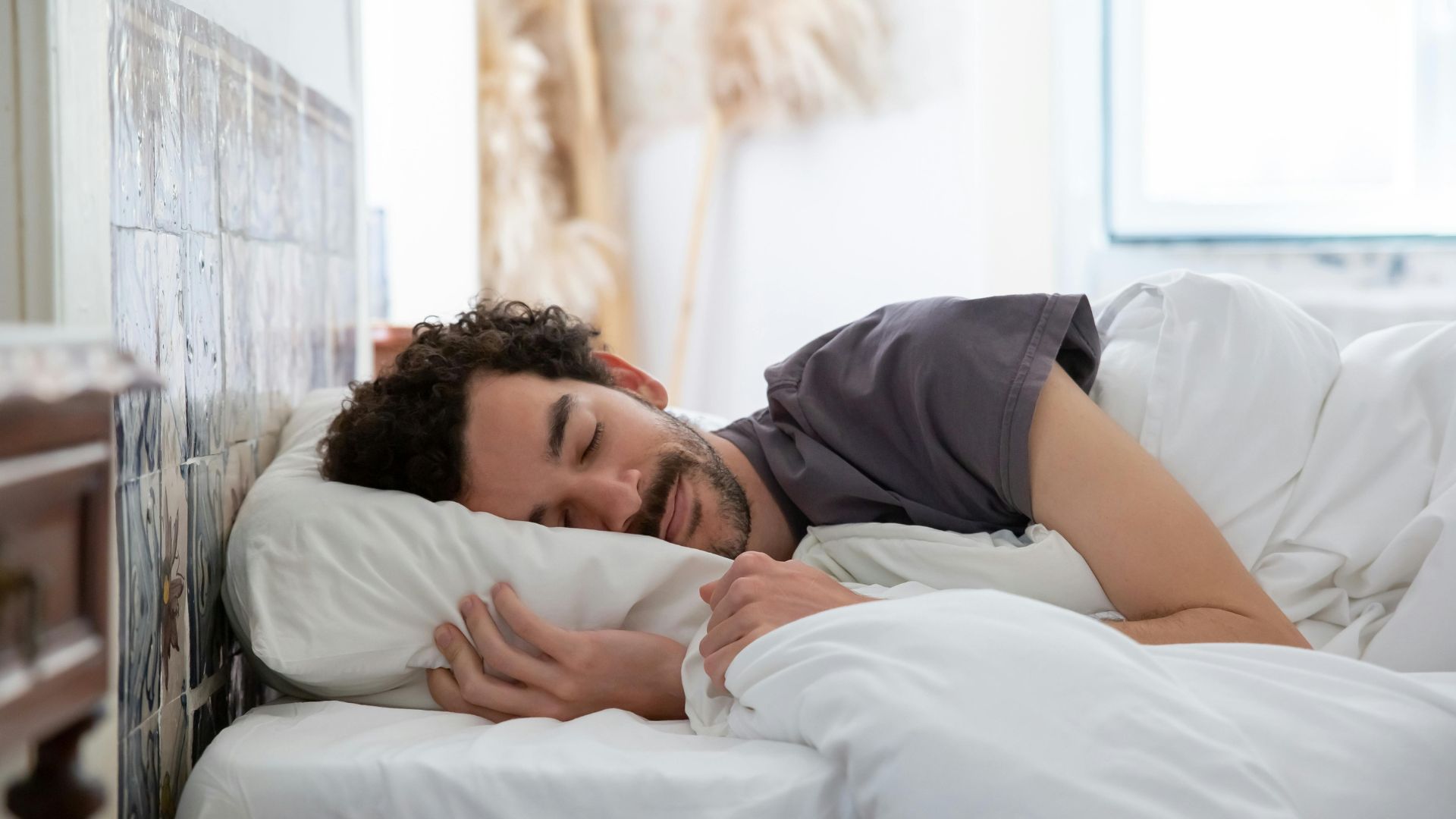
A word about mouth taping
The evidence is thinner than the social media presence. A few small studies in selected mouth-breathing patients show modest improvements (Lee et al., 2022, small cohort). Used carefully, in the right patient, it can be a useful adjunct.
The hard rule: never recommend mouth taping in a patient without confirmed nasal patency. A patient with a blocked nose, taped shut, becomes a patient with obligate oxygen desaturation. The order is always: rule out nasal obstruction → run a Tier 1 trial → only then consider taping for documented persistent mouth-breathing despite an open nose. And always with bed-partner awareness, so someone can intervene if needed.
CLINICAL CORNER
The One-Page PSG → Phenotype Worksheet
For your next OAT consultation, pull the patient’s PSG report and answer four questions. The dominant phenotype is usually obvious by the time you’ve answered all four.
1. Supine AHI vs. lateral AHI? - Supine >2× lateral → Anatomical weighting - Roughly equal → consider non-positional drivers
2. REM AHI vs. NREM AHI? - REM substantially higher (often 2–3×) → Muscle responsiveness and/or loop gain weighting - Roughly equal or NREM-dominant → anatomical or arousal threshold weighting
3. Arousal index relative to AHI? - AI high (>25/hr) with O₂ nadir >85% → Arousal threshold weighting - AI proportional to AHI → arousal threshold less likely the driver
4. Desaturation pattern on the hypnogram? - Cyclical/periodic → Loop gain weighting - Position-locked, resolves laterally → anatomical - Deep, REM-clustered → muscle responsiveness
Document the dominant phenotype and any contributing phenotypes in your treatment note. When you consent the patient for OAT, you’ll have a defensible, evidence-based rationale for combining therapies — and a referral hook for any phenotype that needs sleep-medicine co-management. We’ll build the consent and referral templates in Part 3.
Reading HST output instead of PSG? Most of you are.
The PSG worksheet above is the teaching gold standard. The HST/CPC reality is what most of your patients actually walk in with. Click here for the HST/CPC companion worksheet — same four-question framework, adapted to the data your ring or unattended study actually produces, with SleepImage as the showcase example. Includes what HST data shows clearly, what it shows as a pattern instead of a number, and what it doesn't tell you at all.
COACH CATHY'S TAKE
By Cathy Bennett, MS, NBCHWC
The Tier 1 nasal protocol above is excellent, and it works much better when you remove the thing inflaming the airway tissue in the first place.
In our practice, the patients Mike sees whose noses won't open with saline and xylitol almost always have an underlying inflammatory dietary pattern. The usual suspects: dairy (in the sensitive subset), refined sugar, ultra-processed foods, alcohol within three hours of bed, and chronic dehydration. None of these is causing the OSA. But every one of them keeps the nasal and pharyngeal mucosa swollen, secretory, and reactive, which is exactly the condition that makes an appliance underperform.
A simple six-week test I often suggest, paired with starting Tier 1: cut dairy and added sugar, hit half your bodyweight in ounces of water a day, and protect a 3-hour gap between alcohol and bedtime. Track snoring loudness with a partner or a sleep app. If the nose opens, you've found a lever that contributes, and the appliance has a fairer fight.

It's not glamorous advice. It's the advice that makes the appliance appear to be doing its job.
P.S. The reading list I put together for Issue 1 — the foundational PALM and OAT phenotyping papers from Eckert, Edwards, the AASM/AADSM guideline, and the DISE outcome studies — is [still available here]. Worth bookmarking for any case where you find yourself wondering whether you're looking at an arousal-threshold patient or a true anatomical responder.
Next Week — Part 3
The consent template, the co-management referral letter, and the escalation decision tree. If Part 1 was the science and Part 2 was the clinical workflow, Part 3 is the paperwork that protects the practice and the patient when you do this honestly.
If a sleep medicine colleague has been wondering why your appliance success rate is climbing, this is the issue to forward to them.
REFERENCES
Eckert DJ, White DP, Jordan AS, Malhotra A, Wellman A. Defining phenotypic causes of obstructive sleep apnea: identification of novel therapeutic targets. Am J Respir Crit Care Med. 2013;188(8):996-1004. https://doi.org/10.1164/rccm.201303-0448OC
Eckert DJ, Owens RL, Kehlmann GB, et al. Eszopiclone increases the respiratory arousal threshold and lowers the apnoea/hypopnoea index in obstructive sleep apnoea patients with a low arousal threshold. Clin Sci (Lond). 2011;120(12):505-514. https://doi.org/10.1042/CS20100588
Edwards BA, Sands SA, Eckert DJ, et al. Acetazolamide improves loop gain but not the other physiological traits causing obstructive sleep apnoea. J Physiol. 2012;590(5):1199-1211. https://doi.org/10.1113/jphysiol.2011.223925
Wellman A, Malhotra A, Jordan AS, Stevenson KE, Gautam S, White DP. Effect of oxygen in obstructive sleep apnea: role of loop gain. Respir Physiol Neurobiol. 2008;162(2):144-151. https://doi.org/10.1016/j.resp.2008.05.019
Camacho M, Certal V, Abdullatif J, et al. Myofunctional therapy to treat obstructive sleep apnea: a systematic review and meta-analysis. Sleep. 2015;38(5):669-675. https://doi.org/10.5665/sleep.4652
Strollo PJ, Soose RJ, Maurer JT, et al. Upper-airway stimulation for obstructive sleep apnea. N Engl J Med. 2014;370(2):139-149. https://doi.org/10.1056/NEJMoa1308659
Sutherland K, Phillips CL, Cistulli PA. Efficacy versus effectiveness in the treatment of obstructive sleep apnea: CPAP and oral appliances. J Dent Sleep Med. 2015;2(4):175-181. https://aadsm.org/docs/JDSM.2.4.175.pdf
Hermelingmeier KE, Weber RK, Hellmich M, Heubach CP, Mösges R. Nasal irrigation as an adjunctive treatment in allergic rhinitis: a systematic review and meta-analysis. Am J Rhinol Allergy. 2012;26(5):e119-e125. https://doi.org/10.2500/ajra.2012.26.3787
Camañes-Gonzalvo S, Bellot-Arcís C, Marco-Pitarch R, et al. Comparison of the phenotypic characteristics between responders and non-responders to obstructive sleep apnea treatment using mandibular advancement devices in adult patients: systematic review and meta-analysis. Sleep Med Rev. 2022;64:101644. https://doi.org/10.1016/j.smrv.2022.101644
Lee YC, Lu CT, Cheng WN, Li HY. The impact of mouth-taping in mouth-breathers with mild obstructive sleep apnea: a preliminary study. Healthcare (Basel). 2022;10(9):1755. https://doi.org/10.3390/healthcare10091755
Until next week,
Dr. Michael & Cathy Bennett
More Than Teeth | A Mission for Generational Health
Reply